In this section we will discuss the intracellular components that are not
organelles. The cytoskeleton and cytosol are structural elements
that help provide the cell with its structure. The cytoskeleton is composed of
protein filaments and is found throughout the inside of a eukaryotic cell. The
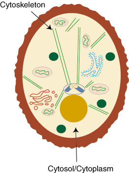
cytosol is the main component of the cytoplasm, the fluid that fills the
inside of the cell. The cytoplasm is everything in the cell except for the
cytoskeleton and membrane-bound organelles. Both structures, the cytoskeleton
and cytosol, are "filler" structures that do not contain essential biological
molecules but perform structural functions within a cell.
The interior of a cell is composed of organelles, the cytoskeleton, and the
cytosol. The cytosol often comprises more than 50% of a cell's volume. Beyond
providing structural support, the cytosol is the site wherein protein
synthesis takes place, and the provides a home
for the centrosomes and centrioles. These organelles will be discussed
more with the cytoskeleton.

The cytoskeleton is composed of three different types of protein filaments: actin, microtubules, and intermediate filaments.
The Cytosol

Figure %: Location of the cytosol within a cell.
The Cytoskeleton
The cytoskeleton is similar to the lipid bilayer in that it helps provide the interior structure of the cell the way the lipid bilayer provides the structure of the cell membrane. The cytoskeleton also allows the cell to adapt. Often, a cell will reorganize its intracellular components, leading to a change in its shape. The cytoskeleton is responsible for mediating these changes. By providing "tracks" with its protein filaments, the cytoskeleton allows organelles to move around within the cell. In addition to facilitating intracellular organelle movement, by moving itself the cytoskeleton can move the entire cells in multi-cellular organisms. In this way, the cytoskeleton is involved in intercellular communication.The cytoskeleton is composed of three different types of protein filaments: actin, microtubules, and intermediate filaments.
Actin
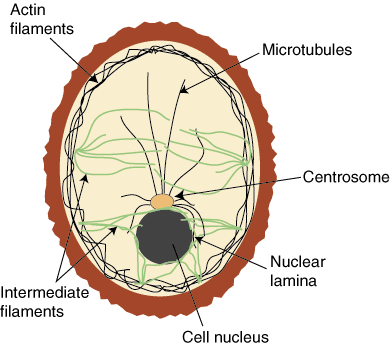
Actin is the main component of actin filaments, which are double-stranded, thin, and flexible structures. They have a diameter of about 5 to 9 nanometers. Actin is the most abundant protein in most eukaryotic cells. Most actin molecules work together to give support and structure to the plasma membrane and are therefore found near the cell membrane.Microtubules
Microtubules are long, cylindrical structures composed of the protein tubulin and organized around a centrosome, an organelle usually found in the center of the cell near the cell nucleus. Unlike actin molecules, microtubules work separately to provide tracks on which organelles can travel from the center of the cell outward.
Microtubules are much more rigid than actin molecules and have a larger
diameter: 25 nanometers. One end of each microtubule is embedded in the
centrosome; the microtubule grows outward from there. Microtubules are
relatively unstable and go through a process of continuous growth and decay.
Centrioles are small arrays of microtubules that are found in the
center of
a centrosome. Certain proteins will use microtubules as tracks for laying out
organelles in a cell.
Intermediate filaments are the final class of proteins that compose the
cytoskeleton. These structures are rope-like and fibrous, with a diameter of
approximately 10 nanometers. They are not found in all animal cells, but in
those in which they are present they form a network surrounding the nucleus
often called the nuclear lamina. Other types of intermediate filaments
extend through the cytosol. The filaments help to resist stress and increase
cellular stability.

These three types of protein are distinct in their structure and specific
function, but all work together to help provide intra-cellular structure.
Because they are so diverse, it is very difficult to study the specific
functions of the cytoskeletal components.
Intermediate filaments

Figure %: Organization of actin, microtubules, and intermediate filaments within
a cell.
No comments:
Post a Comment