Showing posts with label Biology. Show all posts

Wednesday, August 8, 2018

How to Cite This SparkNote

Full Bibliographic Citation

MLA

SparkNotes Editors. “SparkNote on Intracellular Components.” SparkNotes.com. SparkNotes LLC. n.d.. Web. 7 Aug. 2018.

The Chicago Manual of Style

SparkNotes Editors. “SparkNote on Intracellular Components.” SparkNotes LLC. n.d.. http://www.sparknotes.com/biology/cellstructure/intracellularcomponents/ (accessed August 7, 2018).

APA

SparkNotes Editors. (n.d.). SparkNote on Intracellular Components. Retrieved August 7, 2018, from http://www.sparknotes.com/biology/cellstructure/intracellularcomponents/

In Text Citation

MLA

“Their conversation is awkward, especially when she mentions Wickham, a subject Darcy clearly wishes to avoid” (SparkNotes Editors).

APA

“Their conversation is awkward, especially when she mentions Wickham, a subject Darcy clearly wishes to avoid” (SparkNotes Editors, n.d.).

Footnote

The Chicago Manual of Style

Chicago requires the use of footnotes, rather than parenthetical citations, in conjunction with a list of works cited when dealing with literature.
1 SparkNotes Editors. “SparkNote on Intracellular Components.” SparkNotes LLC. n.d.. http://www.sparknotes.com/biology/cellstructure/intracellularcomponents/ (accessed August 7, 2018).

Please be sure to cite your sources. For more information about what plagiarism is and how to avoid it, please read our article on The Plagiarism Plague. If you have any questions regarding how to use or include references to SparkNotes in your work, please tell us.

Problems

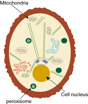
Problem : Identify the cell nucleus, mitochondria, and peroxisomes in the following diagram.

Figure %: Eukaryotic cell


Figure %: Solution
Problem : What is the name of the structures into which DNA is packaged inside the cell nucleus?
DNA is packaged into chromosomes with proteins in the cell nucleus.
Problem : Does the nucleus allow molecules to pass across its double membrane?
Yes. The nuclear membrane contains pores that allow only certain molecules to pass across its membrane either in or out of the cell.
Problem : Why does the mitochondria have two distinct sub-compartments?
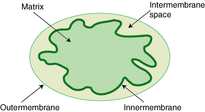
Mitochondrial membranes have two layers—an inner and outer membrane. As a result, there are two distinct spaces in the organelle. One between the inner and outer membranes, called the intermembrane space, and one within the inner membrane, called the matrix space.
Problem : What is the chemical process that occurs in the peroxisomes?
Oxidation. The peroxisomes contain enzymes that oxidize intracellular molecules like fatty acids.
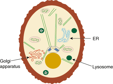
Problem : Identify the endoplasmic reticulum, golgi apparatus, and lysosome in the following diagram.

Figure %: Eukaryotic cell


Figure %: Solution
Problem : Why is it called "rough" endoplasmic reticulum?
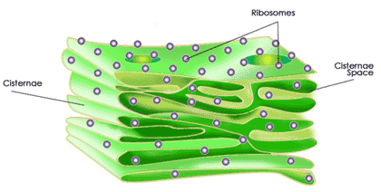
It is called rough endoplasmic reticulum because it is coated with small ribosomal particles that for a bumpy coat on the cytosolic side of the ER membrane. It is contrasted with smooth ER which lacks these ribosomes.
Problem : How are proteins segregated in the golgi apparatus for secretion?
Most proteins contain a signal sequence at its end that tell the golgi to where in the cell it should be secreted.
Problem : Into which face of the golgi apparatus do proteins from the ER enter? Which face do they exit from?
Proteins enter the golgi apparatus through the cis face and exit through the trans face.
Problem : What is the function of the lysosome?
The lysosome functions to remove intracellular debris by digesting it with the acidic enzymes found inside it. 

Eukaryotic Organelles: The Cell Nucleus, Mitochondria, and Peroxisomes

 We will now begin our discussion of intracellular organelles. As we have mentioned, only eukaryotic cells have intracellular sub-divisions, so our discussion will exclude prokaryotic cells. We will also focus on animal cells, since plant cells have a number of further specialized structures. In this section we will discuss the importance of the cell nucleus, mitochondria, peroxisomes, endoplasmic reticulum, golgi apparatus, and lysosome.

The Cell Nucleus

The cell nucleus is one of the largest organelles found in cells and also plays an important biological role. It composes about 10% of the total volume of the cell and is found near the center of eukaryotic cells. Its importance lies in its function as a storage site for DNA, our genetic material. The cell nucleus is composed of two membranes that form a porous nuclear envelope, which allows only select molecules in and out of the cell.
The DNA that is found in the cell nucleus is packaged into structures called chromosomes. Chromosomes contain DNA and proteins and carry all the genetic information of an organism. The nucleus gains support from intermediate filaments that both form the surrounding nuclear lamina and makes direct contact with the endoplasmic reticulum. The nucleus is also the site of DNA and RNA synthesis.

Figure %: Location of the cell nucleus, mitochondria, and peroxisomes in a cell.

Mitochondria

The mitochondria, with its specialized double-membrane structure, generate adenosine triphosphate (ATP), a molecule that provides organisms with energy.

Figure %: Mitochondrial structure
The outer and inner membranes of the mitochondria form two sub-compartments: the internal matrix space and the intermembrane space. Those few proteins found withn the mitochondria are located within the inner membrane. Mitochondria synthesize ATP with energy supplied by the electron transport chain and a process called oxidative phosphorylation.

Peroxisomes

Peroxisomes are single-membrane structures found in all eukaryotic cells. They are small, membrane-bound structures that use molecular oxygen to oxidize organic molecules. The structure is one of the major oxygen utilizing organelles, the other being the mitochondria. Peroxisomes contain oxidative enzymes and other enzymes that help produce and degrade hydrogen peroxide.
Because of their varying enzymatic compositions, peroxisomes are diverse structures. Their main function is to help breakdown fatty acids. They perform specific functions in plant cells, which we will discuss later.

  The Endoplasmic Reticulum

The endoplasmic reticulum, or ER, is a very important cellular structure because of its function in protein synthesis and lipid synthesis. For example, the ER is the site of production of all transmembrane proteins. Since nearly all proteins that are secreted from a cell pass through it, the ER is also important in cellular trafficking. In addition to these major roles, the ER plays a role in a number of other biological processes. There are two different types of ER: smooth ER and rough ER (RER).
The rough ER has its name because it is coated with ribosomes, the structures most directly responsible for carrying out protein synthesis. Smooth ER lacks these ribosomes and is more abundant in cells that are specific for lipid synthesis and metabolism.

Figure %: The Endoplasmic Reticulum
In addtion to protein and lipid synthesis, the ER also conducts post-synthesis modifications. One such modification involves the addition of carbohydrate chains to the proteins, though the function of this addition is unknown. Another major modification is called protein folding, whose name is rather self- explanatory. Another role of the ER is to capture calcium for the cell from the cytosol. Finally, the ER can secrete proteins into the cell that are usually destined for the golgi apparatus.

Figure %: The location of the Endoplasmi Reticulum, golgi apparatus, and lysosome in a eukaryotic cell.

The Golgi Apparatus

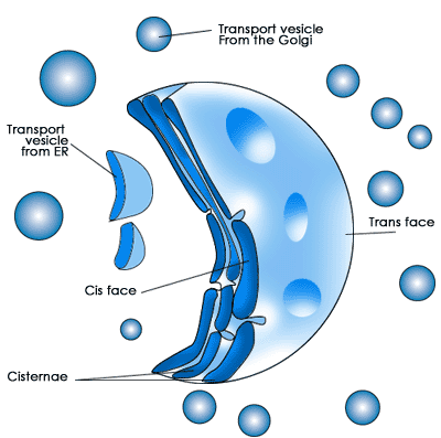
The golgi apparatus is usually located near the cell nucleus. It is composed of a series of layers called golgi stacks. Proteins from the ER always enter and exit the golgi apparatus from the same location. The cis face of the golgi is where proteins enter. A protein will make its way through the golgi stacks to the other end called the trans face where it is secreted to other parts of the cell.

Figure %: Structure of the Golgi Apparatus
In the golgi apparatus, more carbohydrate chains are added to the protein while other chains are removed. The golgi stacks also sort proteins for secretion. After sorting, the membrane of the golgi buds off, forming secretory vesicles that transport proteins to their specific destination in the cell. A protein's destination is often signaled with a specific amino acid sequence at its end. A protein secretion most often travels back to the ER, to the plasma membrane where it can become a transmembrane protein, or to the next structure we will discuss, the lysosomes.

Lysosomes

Lysosomes are sites of molecular degradation found in all eukaryotic cells. They are small, single-membrane packages of acidic enzymes that digest molecules and are found throughout eukaryotic cells. As such, Lysosomes are a sort of cellular "garbage can," getting rid of cellular debris. Proteins that are not correctly folded or have significant mutations can be secreted to the lysosomes and be degraded instead of taking up space in the cell. Detritus proteins and other molecules can find their way to the lysosome in a variey of ways.

Molecules from outside a cell can be taken in through a process called endocytosis. In this process, the cell membrane invaginates, forming a vesicle containing the transported molecule that will eventually reach a lysosome. The reverse of endocytosis is exocytosis. In this process, molecules within a cell are secreted into an endosome, a membrane-bound structure that delivers the molecule to the lysosome. After reaching the lysosomes, the molecules are secreted from a cell in membrane vesicles. Proteins secreted by the golgi apparatus into the plasma membrane can also be taken back to the lysosome by endosomes. 

Problems

Problem : What are the main functions of the cytosol and cytoskeleton?
The cytosol is the site of protein synthesis and the cytoskeleton helps provide intracellular structure and helps with organelle movement in cells.
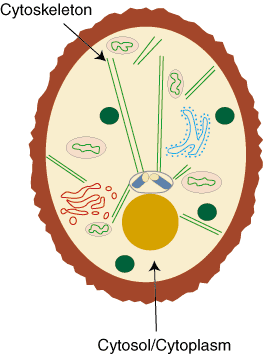
Problem : Label the locations of the cytoskeleton and cytosol in the following diagram.

Figure %: Cell


Figure %: Solution
The cytosol is a liquid that fills the interior of a cell. The cytoskeleton is composed of protein filaments found in the cytosol.
Problem : In what way is the function of the cytoskeleton similar to the function of the lipid bilayer?
The lipid bilayer provides the structure of the cell membrane. Similarly, the cytoskeleton helps provide the structure of the interior of the cell.
Problem : The cytosol makes up most of what intracellular component?
The cytoplasm.
Problem : Name the three proteins that compose the cytoskeleton. Which of these is responsible for forming the nuclear lamina?
The three types of protein filaments are actin molecules, microtubules, and intermediate filaments. Intermediate filaments form the nuclear lamina. 

The Cytoskeleton and Cytosol

 In this section we will discuss the intracellular components that are not organelles. The cytoskeleton and cytosol are structural elements that help provide the cell with its structure. The cytoskeleton is composed of protein filaments and is found throughout the inside of a eukaryotic cell. The cytosol is the main component of the cytoplasm, the fluid that fills the inside of the cell. The cytoplasm is everything in the cell except for the cytoskeleton and membrane-bound organelles. Both structures, the cytoskeleton and cytosol, are "filler" structures that do not contain essential biological molecules but perform structural functions within a cell.

The Cytosol

The interior of a cell is composed of organelles, the cytoskeleton, and the cytosol. The cytosol often comprises more than 50% of a cell's volume. Beyond providing structural support, the cytosol is the site wherein protein synthesis takes place, and the provides a home for the centrosomes and centrioles. These organelles will be discussed more with the cytoskeleton.

Figure %: Location of the cytosol within a cell.

The Cytoskeleton

The cytoskeleton is similar to the lipid bilayer in that it helps provide the interior structure of the cell the way the lipid bilayer provides the structure of the cell membrane. The cytoskeleton also allows the cell to adapt. Often, a cell will reorganize its intracellular components, leading to a change in its shape. The cytoskeleton is responsible for mediating these changes. By providing "tracks" with its protein filaments, the cytoskeleton allows organelles to move around within the cell. In addition to facilitating intracellular organelle movement, by moving itself the cytoskeleton can move the entire cells in multi-cellular organisms. In this way, the cytoskeleton is involved in intercellular communication.
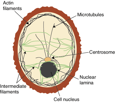
The cytoskeleton is composed of three different types of protein filaments: actin, microtubules, and intermediate filaments.

Actin

Actin is the main component of actin filaments, which are double-stranded, thin, and flexible structures. They have a diameter of about 5 to 9 nanometers. Actin is the most abundant protein in most eukaryotic cells. Most actin molecules work together to give support and structure to the plasma membrane and are therefore found near the cell membrane.

Microtubules

Microtubules are long, cylindrical structures composed of the protein tubulin and organized around a centrosome, an organelle usually found in the center of the cell near the cell nucleus. Unlike actin molecules, microtubules work separately to provide tracks on which organelles can travel from the center of the cell outward.

 Microtubules are much more rigid than actin molecules and have a larger diameter: 25 nanometers. One end of each microtubule is embedded in the centrosome; the microtubule grows outward from there. Microtubules are relatively unstable and go through a process of continuous growth and decay. Centrioles are small arrays of microtubules that are found in the center of a centrosome. Certain proteins will use microtubules as tracks for laying out organelles in a cell.

Intermediate filaments

Intermediate filaments are the final class of proteins that compose the cytoskeleton. These structures are rope-like and fibrous, with a diameter of approximately 10 nanometers. They are not found in all animal cells, but in those in which they are present they form a network surrounding the nucleus often called the nuclear lamina. Other types of intermediate filaments extend through the cytosol. The filaments help to resist stress and increase cellular stability.

Figure %: Organization of actin, microtubules, and intermediate filaments within a cell.
These three types of protein are distinct in their structure and specific function, but all work together to help provide intra-cellular structure. Because they are so diverse, it is very difficult to study the specific functions of the cytoskeletal components. 

Terms

 Actin  -  A very abundant protein in eukaryotic cells that is the main component of actin filaments.
Actin Filaments  -  Approximately 5-9 nanometers in diameter. Provide structural support to the plasma membrane. As a cytoskeletal protein provides for movement of organelles within cells.
Centromere  -  A round structure that holds together sister chromatids.
Centrosome  -  A region of the cell near the nucleus from which microtubules sprout. Centrosomes are not found in all cells. Centrosomes are comprised of two centrioles.
Chromosome  -  A structure composed of DNA and proteins containing all the genetic material of a cell. Found in the cell nucleus.
Cytoplasm  -  A fluid found in the main compartment of eukaryotic cells. Includes everything outside the cell nucleus but the organelles and the cytoskeleton. The main component is cytosol.
Cytoskeleton  -  A system of protein filaments found throughout the cytoplasm of eukaryotic cells that help provide for cell structure. Composed of actin, intermediate filaments, and microtubules.
Cytosol  -  The main component of the cytoplasm that fills the main compartment of eukaryotic cells.
Endoplasmic reticulum  -  A membrane-bound organelle found in eukaryotic cells. Makes direct contact with the cell nucleus and, since it is dotted with ribosomes, is the site of lipid and protein synthesis. Comes in two forms, smooth and rough.
Endosome  -  A membrane-bound organelle found in eukaryotic cells. Responsible for delivering molecules to the lysosome for digestion.
Eukaryote  -  An organism composed of one or more cells with defined intracellular components including a nucleus and cytosol. Includes all organisms except bacteria and viruses.
Golgi apparatus  -  A membrane-bound organelle found near the cell nucleus in eukaryotic cells. Responsible for sorting and packaging proteins for secretion to various destinations in the cell.
Intermediate filament  -  One of three protein components of the cytoskeleton. A fibrous protein filament approximately 10 nanometers in diameter. Forms the nuclear lamina that helps protect the cell nucleus.
Intermembrane space  -  The space between the outer and inner membrane in a mitochondria.
Lysosome  -  A membrane-bound organelle found in eukaryotic cells. Contain acids and enzymes that degrade unwanted molecules.
Matrix  -  The space inside the inner membrane of mitochondria.
Microtubule  -  One of three protein components of the cytoskeleton. Long, cylindrical structures approximately 25 nanometers in diameter. Extend from the centrosome to all parts of the cell, forming tracks on which organelles can travel within the cell. Microtubules can be either kinetocore microtubules or non-kinetocore microtubules. Kinetocore microtubules bind to sister chromatids during mitosis; non-kinetocore microtubules do not.
Mitochondria  -  An organelle within the cell. Much of cell respiration is carried out within its bounds.
Nucleus  -  A large, double membrane-bound organelle found in eukaryotic cells. Contains DNA and RNA.
Organelle  -  A membrane-bound sub-cellular structure found in eukaryotic cells. The Cell nucleus, mitochondria, ER, and golgi apparatus are all examples.
Peroxisome  -  A small, membrane-bound organelle found in eukaryotic cells. Contains oxidizing enzymes that oxidize organic molecules and process hydrogen peroxide in the cell.
Prokaryote  -  An organism composed of usually one, but occasionally more, cells that lack defined sub-cellular compartments. All essential material is enclosed within the cell membrane. Includes all bacteria and close relatives.
Ribosome  -  A molecule composed of ribosomal RNA* {biology/molecularbiology/translation}* and proteins, and located on the endoplasmic reticulum**. Responsible for mediating protein synthesis.
Rough endoplasmic reticulum  -  Endoplasmic reticulum that is coated with ribosomes and involved in protein synthesis.
Smooth endoplasmic reticulum  -  Naked endoplasmic reticulum that lacks ribosomes and is more involved in lipid synthesis. 

Introduction to Intracellular Components

 Now that we have discussed the one universal structural element of cells, the cell membrane, we will begin reviewing the specific intracellular components found in eukaryotes, or multi-cellular organisms. Eukaryotes differ from prokaryotes in the level of their structural complexity. Whereas the simpler prokaryotes contain all their genetic material, such as DNA and RNA, within the cell membrane, eukaryotes have intracellular compartments enclosing different structures, called organelles, which contain different molecules and enzymes that perform various functions within and between cells.
In this section, we will discuss the function, structure, and location of intracellular compartments found in eukaryotic cells. With an understanding of these principles we can see how these components are necessary for cell life. While eukaryotic cells have increased structural complexity, prokaryotic cells are able to carry out most of the same processes with their simple structure. We will discuss what significance the different components have in eukaryotic cell life.
We will begin our discussion of eukaryotic intracellular components by discussing the structural roles of the cytoskeleton and cytosol. We will then discuss the biological function of various organelles, including the cell nucleus, mitochondria, peroxisome, endoplasmic reticulum, golgi apparatus, lysosome, endosome, and related structures. The nucleus and mitochondria house DNA and provide the cell with energy, respectively. Peroxisomes and lysosomes are responsible for degrading molecules within the cell. The endoplasmic reticulum, golgi apparatus, and endosomes are involved in cellular transport. 

Cell Differences

Introduction to Intracellular Components
Terms

Summary and Analysis


How to Cite This SparkNote

Problems

Problem : Why is it necessary for cell membranes to have proteins that help transport molecules?
The cell membrane is composed of a lipid bilayer that is highly impermeable to most molecules. As a result, outside structures are required to help transport essential large, polar molecules across the cell membrane.
Problem : What is the name of the natural process by which molecules flow from an area of higher concentration to one of lower concentration?
Diffusion.
Problem : What is the difference between the behavior of carrier and channel proteins?
Carrier proteins allow specific molecules to cross the cell membrane by undergoing a conformational change upon the binding of the molecule. The conformational change opens a hole through which the molecule can enter or exit a cell. Channel proteins do not require the binding of a molecule and conformational change to open. Channel proteins allow molecules to steadily diffuse across the membrane through diffusion.
Problem : What is the function of an ionophore?
Ionophores function to increase a membrane's permeability to a specific ion thereby facilitating its movement across that cell's membrane.
Problem : Name two specific functions of membrane transport.
Membrane transport helps maintain the proper distribution of ions across a cell membrane; helps maintain proper cellular pH, and helps mediate communication between cells in multi-cellular organisms. 

Structures Responsible for Membrane Transport

 We have discussed how the lipid bilayer acts as an efficient barrier by only allowing a very small number of non-polar molecules to freely enter or exit a cell. While for the most part this selectivity is a valuable function and allows the cell to maintain its integrity, cells do need to move certain large, polar molecules such as amino acids, sugars, and nucleotides across their membranes. As a result, cell membranes require specific structures that allow for the transport of certain molecules.

Membrane Transport

There are a number of different ways that molecules can pass from one side of a cell membrane to the other. Some such means, like diffusion and osmosis, are natural processes that require no expenditure of energy from the cell and are called passive transport. Other methods of transport do require cellular energy and are called active transport. In addition to these two forms of transport, there exist other forms of transport such as endocytosis and exocytosis, which will be discuss later and do not require the same set of membrane proteins for their function.

Passive Transport

Diffusion is the natural phenomenon in which nonpolar molecules naturally flow from an area of higher concentration to an area of lower concentration. Osmosis is a similar process, but refers specifically to water molecules. Both of these classes of molecules we have already discussed as capable of crossing the lipid bilayer. As seen in , neither diffusion nor osmosis require the expenditure of energy.

Active Transport

Active transport occurs when a cell actively pumps a molecule across its membrane, against the natural direction dictated by diffusion, osmosis, or polarity. As seen in , such transport requires energy.

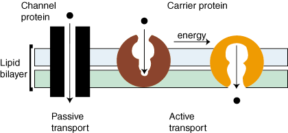
Figure %: Active and Passive Transport Proteins

Transport Proteins

Both of passive and active transport are mediated with the help of transmembrane proteins that act as transporters. shows the two main classes of transport proteins: carrier proteins and channel proteins. For the most part, carrier proteins mediate active transport while channel proteins mediate passive transport. Carrier proteins create an opening in the lipid bilayer by undergoing a conformational change upon the binding of the molecule. Channel proteins form hydrophilic pores across the lipid bilayer. When open, these pores allow specific molecules to pass through. There is one other class of transport proteins called ionophores. These are small, hydrophobic proteins that increase bilayer permeability for specific ions.
Transport proteins are critical to cell life and cell interactions. They allow for the proper distribution of ions and molecules in multicellular organisms. Additionally, they can help to maintain proper intra- and extra-cellular pH levels, facilitate communication between cells, and are involved numerous other essential functions including protein sythesis. 

Problems

Problem : Will you find the same set of membrane proteins in each cell membrane?
No. Membrane proteins perform a number of functions within cells, as a result, different proteins are necessary in different regions of cells depending on the function of the cell and the interactions it may take part in.
Problem : What are the names of the two main classes of membrane proteins and how could you tell one from the other?
The two main classes of membrane proteins are integral versus peripheral proteins. Since peripheral proteins are easily dissociated from the lipid bilayer, one could treat a cell with a mild detergent that does not disrupt the cell membrane and then see if the specific protein remains associated with the lipid bilayer or is removed.
Problem : What is the name of the configuration that membrane proteins adopt in regions that span the lipid bilayer?
This configuration is called an alpha-helix. It is the same structure that DNA adopts naturally.
Problem : Which class of proteins, integral or peripheral, are freer to move around within the lipid bilayer?
Integral proteins can be thought of as icebergs that float in a lipid bilayer sea. They are relatively mobile in the cell membrane.
Problem : The cell surface is covered with an additional set of molecules. What name is given to these structures and what is their function in the cell?
The cell surface is covered with a cell coat or glycocalyx which consists of carbohydrate chains. They help protect the cell from damage.  

Membrane Proteins

 In addition to the lipid bilayer, the cell membrane also contains a number of proteins. We have already mentioned the presence of certain proteins in the cell membrane. In this section we will discuss the different classes of proteins found there. While the lipid bilayer provides the structure for the cell membrane, membrane proteins allow for many of the interactions that occur between cells. As we discussed in the previous section, membrane proteins are free to move within the lipid bilayer as a result of its fluidity. Although this is true for most proteins, they can also be confined to certain areas of the bilayer with enzymes. Membrane proteins perform various functions, and this diversity is reflected in the significantly different types of proteins associated with the lipid bilayer.

Classifications of Membrane Proteins

Proteins are generally broken down into the smaller classifications of integral proteins, peripheral proteins, and lipid-bound proteins.

Integral Proteins

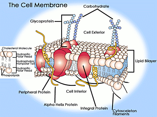
Integral proteins are embedded within the lipid bilayer. They cannot easily be removed from the cell membrane without the use of harsh detergents that destroy the lipid bilayer. Integral proteins float rather freely within the bilayer, much like oceans in the sea. In addition, integral proteins are usually transmembrane proteins, extending through the lipid bilayer so that one end contacts the interior of the cell and the other touches the exterior. The stretch of the integral protein within the hydrophobic interior of the bilayer is also hydrophobic, made up of non-polar amino acids. Like the lipid bilayer, the exposed ends of the integral protein are hydrophilic.

Figure %: Membrane Proteins
When a protein crosses the lipid bilayer it adopts an alpha-helical configuration. Transmembrane proteins can either cross the lipid bilayer one or multiple times. The former are referred to as single-pass proteins and the later as multi-pass proteins. As a result of their structure, transmembrane proteins are the only class of proteins that can perform functions both inside and outside of the cell.

Peripheral Proteins

Peripheral proteins are attached to the exterior of the lipid bilayer. They are easily separable from the lipid bilayer, able to be removed without harming the bilayer in any way. Peripheral proteins are less mobile within the lipid bilayer.

Lipid-Bound Proteins

Lipid-bound proteins are located entirely within the boundaries of the lipid bilayer.

  The Cell Surface

The protein and lipid cell membrane is covered with a layer of carbohydrate chains on its outer surface. This layer is called a cell coat or glycocalyx. The exact composition and distribution of these chains is very diverse. The chains are thought to provide the cell with protection against damage. Glycocalyx are only found on the surface of the cells of higher organism's.

Figure %: A detailed view of a Cell Membrane (phospholipid bilayer and associated proteins) 

Problems

Problem : Identify the lipid bilayer in the following diagram of a cell.

Cell


Solution
The lipid bilayer is the outer-most layer surrounding the cell.
Problem : What is the main function of the lipid bilayer?
The lipid bilayer acts as a barrier between the inside and outside of the cell. It is highly impermeable and does not allow most molecules to freely pass through it into or out of the cell.
Problem : Why is the structure called a lipid bilayer?
It is called a lipid bilayer because it is composed of two layers of fat, or lipid, molecules.
Problem : Fill in the blanks.
A phospholipid molecule contains two distinct regions. The __________ region is attracted to water and the ___________ region is repelled from water. As a result of its both polar and nonpolar regions, it is classified as a(n) __________ molecule.
Hydrophilic. Hydrophobic. Amphipathic.
Problem : Are the two layers of the lipid bilayer identical in composition?
No. As a result of the bilayer's fluidity, structures such as lipids and proteins can freely move around within the lipid bilayer. 

The Lipid Bilayer

  Lipid Bilayer Structure

The lipid bilayer is a universal component of all cell membranes. Its role is critical because its structural components provide the barrier that marks the boundaries of a cell. The structure is called a "lipid bilayer" because it is composed of two layers of fat cells organized in two sheets. The lipid bilayer is typically about five nanometers thick and surrounds all cells providing the cell membrane structure.

Lipids and Phospholipids

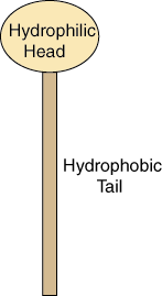
The structure of the lipid bilayer explains its function as a barrier. Lipids are fats, like oil, that are insoluble in water. There are two important regions of a lipid that provide the structure of the lipid bilayer. Each lipid molecule contains a hydrophilic region, also called a polar head region, and a hydrophobic, or nonpolar tail region.

Figure %: Basic Lipid Structure
The hydrophilic region is attracted to aqueous water conditions while the hydrophobic region is repelled from such conditions. Since a lipid molecule contains regions that are both polar and nonpolar, they are called amphipathic molecules.
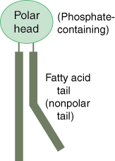
The most abundant class of lipid molecule found in cell membranes is the phospholipid. The phospholipid molecule's polar head group contains a phosphate group. It also sports two nonpolar fatty acid chain groups as its tail.

Figure %: Phospholipid Structure
The fatty acid tail is composed of a string of carbons and hydrogens. It has a kink in one of the chains because of its double-bond structure.

The Bilayer

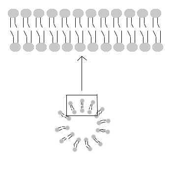
The phospholipids organize themselves in a bilayer to hide their hydrophobic tail regions and expose the hydrophilic regions to water. This organization is spontaneous, meaning it is a natural process and does not require energy. This structure forms the layer that is the wall between the inside and outside of the cell.

Figure %: Lipid Bilayer

Properties of the Lipid Bilayer

As we have already mentioned, the most important property of the lipid bilayer is that it is a highly impermeable structure. Impermeable simply means that it does not allow molecules to freely pass across it. Only water and gases can easily pass through the bilayer. This property means that large molecules and small polar molecules cannot cross the bilayer, and thus the cell membrane, without the assistance of other structures.
 Another important property of the lipid bilayer is its fluidity. The lipid bilayer contains lipid molecules, and, as we will discuss later, it also contains proteins. The bilayer's fluidity allows these structures mobility within the lipid bilayer. This fluidity is biologically important, influencing membrane transport. Fluidity is dependent on both the specific structure of the fatty acid chains and temperature (fluidity increases at lower temperatures).
Structurally, the lipid bilayer is asymmetrical: the lipid and protein composition in each of the two layers is different. 

Terms

 Active transport  -  The transport of molecules across a membrane and against their natural flow; mediated by carrier proteins and requiring outside energy.
Carrier protein  -  A protein responsible for mediating the active transport of molecules from one side of the lipid bilayer to the other. Transport is carried out by a conformational change that occurs within the protein that forms an opening for specific molecules to pass through.
Channel protein  -  A protein responsible for mediating the passive transport of molecules from one side of the lipid bilayer to the other. Transport is carried out by its membrane-spanning hydrophilic structure which, when open, allows molecules to pass through.
Diffusion  -  The transport process in which molecules naturally travel from an area of higher concentration to an area of lower concentration.
Glycocalyx  -  A layer of carbohydrates that coats the exterior of higher-ordered cells. Functions in protecting the cell from damage.
Hydrophilic  -  A polar molecule that selectively associates itself with water through hydrogen bonds.
Hydrophobic  -  A nonpolar molecule that does not readily associate with water through hydrogen bonds.
Integral protein  -  A membrane protein that cannot be easily removed from the lipid bilayer.
Ionophore  -  A class of membrane transport proteins. Small, hydrophobic molecules that increase membrane permeability to certain ions.
Lipid bilayer  -  A thin double layer of phospholipid molecules. Provides the structure of a cell membrane. Structure is a result of hydrophobic and hydrophilic forces.
Lipid-bound protein  -  Membrane proteins that are located entirely within the lipid bilayer, having no part touching either the inside or outside of the cell.
Multi-pass protein  -  Transmembrane proteins that cross the lipid bilayer more than one time.
Osmosis  -  The process by which water naturally travels from an area of high concentration to one of lower concentration.
Passive transport  -  Transport mediated by channel proteins. The movement of molecules across a membrane according to the natural flow.
Peripheral protein  -  A membrane protein that can be easily removed from the lipid bilayer.
Single-pass protein  -  A transmembrane protein that only crosses the lipid bilayer one time.
Transmembrane protein  -  A membrane protein that spans the lipid bilayer having portions in contact with both the inside and outside of the cell. Area within the lipid bilayer forms an alpha-helix.  

Introduction

 Cell membranes are very important structures to cells because they function as a barrier between the components of the cell and the outside environment. The cell membrane is not only responsible for creating a wall between inside and outside the cell, it must also act as a threshold through which select molecules can enter and exit the cell when necessary. The cell membrane is what defines the cell and keeps its components separate from outside cells or organisms.
The cell membrane is composed of a double layer of fat cells called a lipid bilayer in which membrane proteins are embedded. The structure of the lipid bilayer prevents the free passage of most molecules into and out of the cell. We will begin our discussion of the structure of the cell membrane by discussing the structure and properties of the lipid bilayer. We will then go on to discuss the role of membrane-bound proteins, and finally, will discuss membrane transport structures. 

Cell Differences

Introduction
Terms

Summary and Analysis


Introduction to the Cell

 The purpose of this guide is to provide an overview of the basic structural components of living cells. In reviewing these structures, we will also discuss their functions.
All living organisms are composed of cells. A cell is a small, membrane-bound compartment that contains all the chemicals and molecules that help support an organism's life. An understanding of the structure of cells is one of the first steps in comprehending the complex cellular interactions that direct and produce life.
Cells can be thought of as building blocks of organisms. Some organisms are composed of a single cell. Others, like ourselves, are composed of millions of cells that work together to perform the more complex functions that make us different from bacteria. It is difficult to imagine that humans are descendants of a single cell, but this is a common belief in the scientific world. Before we can understand how multiple cells can work together to create complex biological functions, it is necessary to understand what biological functions single cells are capable of performing on their own to sustain life.
There are different types of cells with individuated structures. Single-celled organisms have different cell structure than multi-celled organisms and plant cells have different structures from animal cells. These differences reflect differences in the functions that each of these classes of cells is required to perform. While the focus of this guide will be on the structures that compose complex multi-cellular organisms, we will begin our discussion of cell structure with a structure that is universal to all cells, membranes.